Vassi Talampassi awoke, hazy from anesthesia, in a hospital bed. She pulled her arm from beneath a thin blanket, waving it through the air like a wand. Before a breast cancer diagnosis derailed her life, she’d been an artist and furniture refinisher. The diagnosis had taken so much; she wanted to make sure her lumpectomy and lymph node removal hadn’t taken away her ability to paint.
Talampassi had been diagnosed with stage two invasive lobular carcinoma, a kind of breast cancer that grows in jagged lines, rather than lumps, making it tough to detect in screenings like mammograms. Her mom had survived breast cancer years before and, as Talampassi was wheeled into surgery that afternoon in 2021, she felt safe. She thought about her children, about God, about how she believed the Archangel Michael was protecting her. “I remember thinking, ‘I made the right decision. They will take it out now. I’ll be fine,’” the Windsor, Ont., mother of two says. “I trusted my doctor.”
Ten days later, she sat in her surgeon’s office for what she thought was a routine followup to check on how her scar was healing. Instead, he told her she had something called positive margins from surgery. She was confused.
He explained that the MRI used to locate cancerous tumours during surgery is like a map, useful as a guide but imperfect at a cellular level. For days, Talampassi had been awash with relief that she was cancer-free. Now, the doctor she’d trusted was telling her she still had the disease; microscopic cells had evaded his surgical tools and were floating, sinister and rampant, in her body. Her face flushed with anger. It felt like emotional whiplash.
“What do you mean?” she asked, frantic, thinking of all the testing and scans she’d endured before surgery. “How did this happen?”
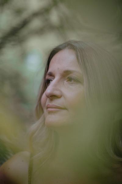
Three weeks later, she was wheeled into another surgery. This time, she was rigid with fear. “What if I don’t wake up?” she thought. She never once considered that she’d again come out of surgery with positive margins.
Two weeks later, she sat in the same surgeon’s office listening to him, feeling like she was trapped in a nightmare. The news that she still had positive margins landed like a hammer blow. “I didn’t know what to do,” she says. “I was so angry.”
In an instant, her mind ran through all the things this news made certain. She’d need a third surgery. She’d require four rounds of chemo. She’d have to finally tell her kids she had cancer. She’d come face to face, again, with the prospect of her own mortality. “I wasn’t ready to die,” she says. “I’m not ready, I’m not there.”
She told her oncologist she was opting to have a double mastectomy. She refused to have that margin conversation again.
After a lumpectomy – breast-conserving surgery to remove a cancerous tumour – surgeons often tell patients they think they got it all. But the reality is that about one in five surgeries doesn’t go that way. And because cancer cells are microscopic, there’s no way to know for sure until a pathology lab can stain, test and examine a tissue sample from the tumour. It’s a process that can take a week or more, leaving patients grasping at hope and comfort as they stare down some of the longest hours of their lives, waiting to learn if they still have cancer or not, and what the rest of their gruelling journey through treatment will look like.
For years, companies have been trying to figure out how to bridge this gap. And while Talampassi sat in her doctor’s office, reeling, a team at a Toronto-based company called Perimeter Medical Imaging AI was working on a possible solution.
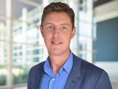
Andrew Berkeley didn’t know much about breast cancer beyond pink ribbons and headscarves. But as an engineer with a background in biophotonics (harnessing light-based tech for medical uses), he suspected he could help. Berkeley worked with a technology called optical coherence tomography, or OCT for short. OCT uses reflected light to capture a portrait of what’s happening under the skin’s surface. The tech was developed by a team at Massachusetts Institute of Technology (MIT) in the early 1990s. Berkeley originally used it in his work at an optometry startup called Tornado Spectral Systems before realizing it could likely do more than eye exams. He and others at the company thought the tech could be used in cancer cases, especially breast cancer, which affects an estimated 32,400 Canadian women a year – and has one of the highest re-excision (or repeat surgery) rates.
In 2013, Berkeley co-founded a new company called Perimeter Medical with a singular focus: create a device that can solve the problem of repeat surgeries for breast cancer. There was a business case for reducing the number of surgeries: one study, published in Current Oncology in 2016, argued that hospitals could save about $1,000 per patient if the re-excision rate dropped from 23 per cent to 10 per cent, leading to $1.9 million of savings annually in the province of B.C. alone. There are other benefits, too: Perimeter’s technology could reduce patients’ emotional toll, making it a rarity in the medical research world, where human experience can often come second to physical outcomes.
Get our weekly newsletter – the people, places, and ideas revealing where Canada is headed.
Berkeley and his co-founder, David Rempel, who’d worked with him as a research lead at Tornado, believed they had the technology to do it. What they needed were samples – as many as they could get – to hone their tech. The pair partnered with Toronto’s Princess Margaret Cancer Centre, one of the world’s leading cancer research hospitals, spending weeks trudging through the snow from their office on Richmond Street West to the basement of the hospital, where they tested their imaging technology on animal and human specimens. They spent years comparing results. Was the camera picking up detail with enough granularity? Did its results match the results the samples had received from pathology? And could it get those results quickly enough to be useful in the operating room?
One afternoon in 2018, Berkeley held two images up side by side. “That’s when the real light bulbs went off. We were able to say, ‘What we see in our images matches what the pathologist looks at under the microscope,’” he says. “I was like, ‘We can do this. We can prove it.’”
The tech was picking up microscopic disease that MRI and ultrasound couldn’t see. Depending on tissue density, the near-infrared light was reflecting (from dense structures like calcifications), transmitting through (from fat or cysts) or scattering (from fibrous tissue). It turned those responses into cellular resolution.
“The question became, ‘Okay, how do we turn this into a scalable product that we can actually get into the operating room?’” It needed to be precise, and it needed to be fast. “Every second counts,” Berkeley says. “You don’t want patients under anesthetic for longer than they need to be.”
Performing even a routine lumpectomy presents a unique challenge: how do you completely remove a tumour you can’t see or feel, while leaving the patient with a cancer-free body they still recognize? It’s an especially difficult question if the patient on the table has a subtype of cancer like ductal carcinoma in situ (DCIS) or invasive lobular carcinoma, whose cells grow in diffuse patterns or spindly lines, like treacherous microscopic veins running through tissue. Compounding the problem is that, unlike a kidney, colon or lung, breasts don’t have defined boundaries or inherent margins. A surgeon removing a kidney tumour takes the whole kidney. Colon surgeons take a bowel segment. In a breast-conserving surgery, margins are defined only by where a surgeon chooses to cut.
Over the last 20 years, surgeons have used a slew of methods to assess whether they have, indeed, gotten it all. Specimen radiography (X-raying the excised tumour to make sure the lesions are centred), can help. So can intraoperative ultrasound or 3D mammography. But the reality is there’s no way in the operating room to be sure. It isn’t until the pathologist returns a finding weeks later, after inking each side of a tumour to assess if any cancer remains, that a patient and their surgeon can truly exhale.
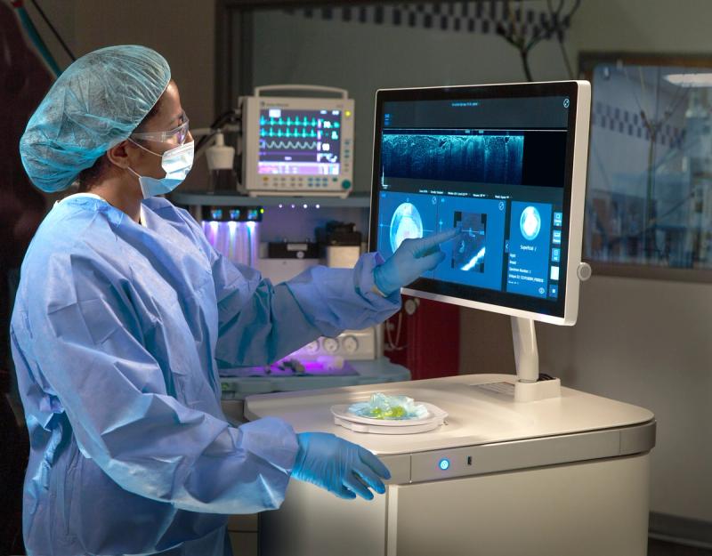
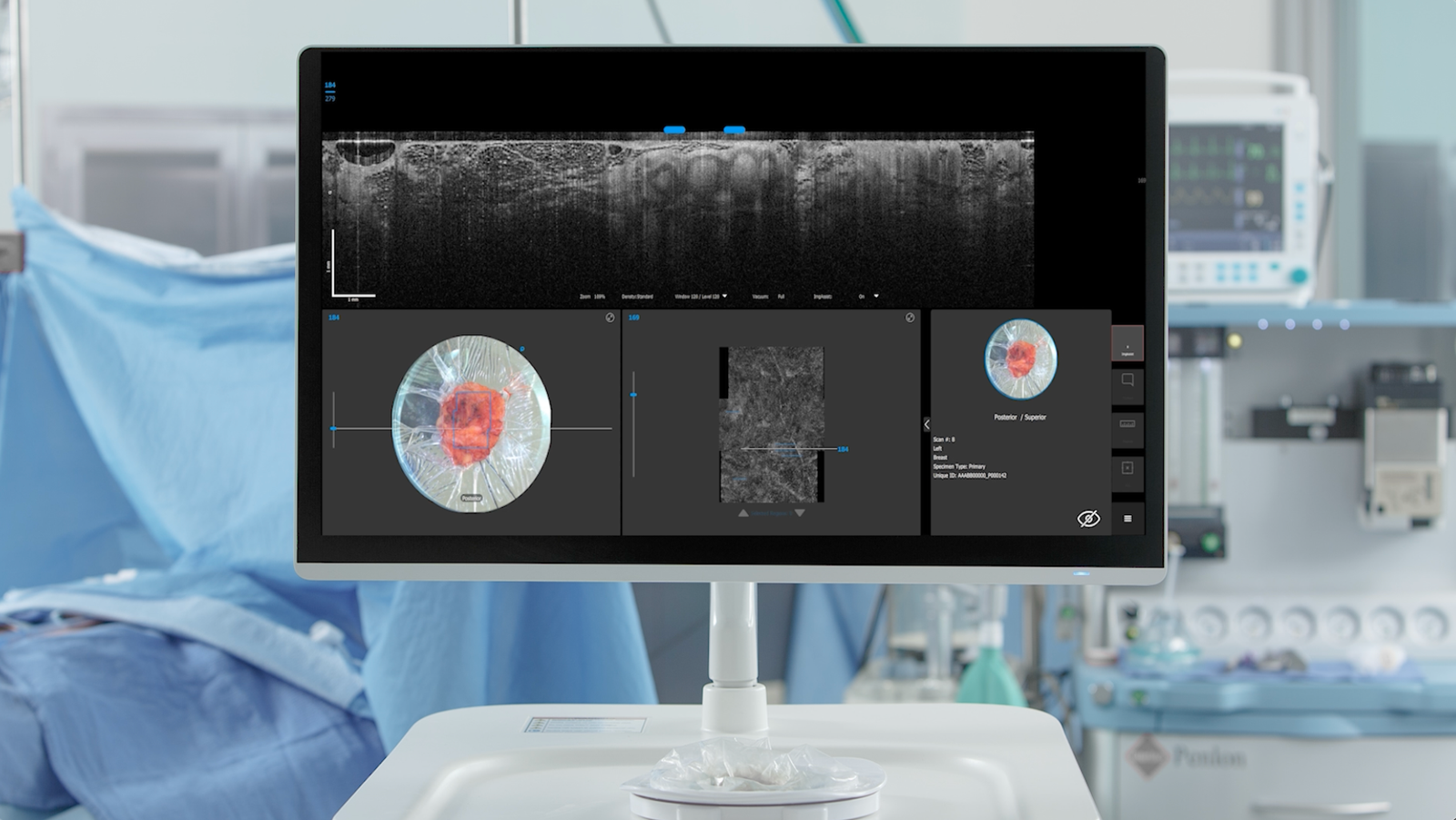
Claire, Perimeter’s imaging device, aims to compress – and one day eliminate – that wait. A device 12 years in the making, Claire is a little like a technological layer cake. On the bottom is the computer, the device’s engine. Atop that is a camera, then a vacuum, then a glass tray. The proverbial cherry on top is a monitor that shows granular imaging with resolution 10 times clearer than an X-ray and 100 times better than an MRI.
Today, a doctor can remove a tissue sample from a patient and place it on a specimen tray that’s unwrapped and slotted into position for each new sample. A vacuum suctions it onto the glass tray, ensuring fewer air bubbles and less movement. The camera begins to scan it from every angle, taking one to two minutes per side. As the images populate, an AI assistant scans them for any suspicious calcifications, flagging areas of interest in red for the surgeon to assess. If cancer cells still exist, the surgeon can go back in and remove them while they’re still in the operating room.
The engineering lab of Perimeter’s Toronto office is serious and playful in equal measure. Shelves are lined with tubes of ultrasound gel and spare keyboards. Pegboards of power tools hang on one wall alongside bins of nuts and washers. Next to an earlier model of Claire that looks like a blue bank safe on wheels is a utility cart with a rainbow of paint tubes, for a project that software engineer Sacha Pompeu-Robinson isn’t at liberty to discuss.
Pompeu-Robinson joined the company in 2015, around the time Perimeter was developing partnerships with Mount Sinai Hospital in New York City and University of Texas MD Anderson Cancer Center in Houston. Once, she carted an earlier iteration of Claire from the manufacturer’s office in Victoria, B.C., to a meeting in Vancouver, loading it into a van, riding with it on a ferry and taking it up an elevator before demonstrating it to investors. Despite the jostling, “it stayed calibrated the whole time,” she says.
Perimeter has landed some major backers, including Social Capital, a firm run by former Facebook executive Chamath Palihapitiya, which proffered a $43.4-million investment. When Palihapitiya learned that his old college friend Adrian Mendes, who’d recently helped build cloud infrastructure company Groq, was casting about for his next challenge, he suggested Perimeter. Mendes is an AI guy and was intrigued by the library of hundreds of thousands of images the company had amassed. It was a data set from a camera that can take pictures no one else can take, and the possibility to use it to train AI on images from the top cancer centres on the continent was too good to pass up.
Mendes arrived as CEO in 2023, at an inflection point for the company. Perimeter was in the midst of a clinical trial, the results of which would determine whether they could apply for U.S. Food and Drug Administration (FDA) approval – the mark of legitimacy for any medical technology company hoping to enter the U.S. market. The trial looked at 206 breast cancer patients, 35 of whom had positive margins after surgery. Claire was able to identify residual cancer in 14 of them, or 40 per cent. It also flagged cancer in six other patients that standard care and the pathology lab hadn’t caught. The trial found Claire could identify with 88.1 per cent accuracy whether margins were clear – not perfect, but a leap forward, nonetheless. The team was thrilled. It was enough to take to the FDA and, hopefully, the market. They began to prepare their application.
The team knew there were other devices on the market, such as one that used a probe to flash red or green and another that used fluorescence to alight if cancer was detected, but none that used OCT or gave a surgeon an AI algorithm that could analyze hundreds of images in real time. They wanted to be the first.
As afternoons dragged into evenings, the team ran through vulnerabilities they needed to address, gaming out all the things the FDA could find fault with and coming up with solutions. What if their AI was out of date by the time the agency examined it? What if their manufacturing supply chain broke down? What if they got an adjudicator who wanted only paper copies of their application, when they’d prepared everything on USB? In the wake of many, many boxes of pizza, they made two sets, analog and digital, just in case.
Mendes was on the phone with an analyst in March 2026 when he got the news of the FDA approval and had to work to stay poised and not reveal what was still a confidential development. He hung up and immediately called the team to celebrate. “We had a sigh of relief, and then the next day, or maybe even the next hour, it’s, ‘Okay, time to run a million miles an hour again,’” he says.
To have a finding come back positive for remaining cancer and to have to deliver that news to a patient is, in a word, terrible, says Dr. Michael Reedijk, senior scientist and surgical oncologist at UHN.
“When you speak to the patient or family member immediately after surgery, they almost always ask you, ‘Did you get it all out?’ And then, you know, of course you say, ‘Yes, I think I got it all out,’ and that’s what sticks with them,” he says.
To learn weeks later that that didn’t happen comes with crushing disappointment and uncertainty. “Patients are also aware that if they need a repeat lumpectomy, that delays the next phase of their treatment, which could be radiation or chemotherapy. There are major, major impacts on patients when you have to revise margins. It can’t be overstated.”
There’s no exact number, but based on the approximately 18,000 to 20,000 lumpectomies performed in Canada per year, at a 15 per cent to 20 per cent national re-excision rate, that’s between 2,700 and 4,000 annual surgeries a device like Claire could prevent.
Reedijk hasn’t seen Claire in action but says he’s cautiously optimistic about the promise of devices like it. “If it’s a technology that fits into the workflow of the operating room, and it’s not prohibitively expensive, it would be extremely welcome and would completely change the way that we manage patients intraoperatively.”
A few months ago, Mendes stood by a demo table at a conference talking to a surgeon. Fifty years ago, the doctor told Mendes, surgical infections were commonplace. They happened all the time, and you just dealt with it. These days, that would be unacceptable, the doctor said. “This is what we need to do with re-operation rates for cancer surgeries, make it so that this is just unacceptable,” Mendes says.
Claire is only the beginning; the company’s version of iPhone v.1, he says. “It’s taking a problem that we’ve all lived with, that our parents lived with, our grandparents lived with, and saying, ‘Our kids won’t have to live with this, because it’ll be totally unacceptable for it to happen anymore.’ That’s what I think we can do.”
Perimeter is already working on devices that would use OCT on other forms of cancer and hopes to close the gap between biopsy and diagnosis, too, a process that can mean weeks of excruciating waiting. The company also hopes to see the device in Canadian hospital rooms and plans to begin the Health Canada approval process after establishing a foothold in the U.S. The FDA approval means the team can start selling Claire to U.S. hospitals; two are already testing it.
Vassi Talampassi will be watching closely. Today, when she speaks to other women facing similar cancer diagnoses, she sees the fear in their eyes.
“If there is a device that is going to save you the emotional pain and the one million thoughts that go on inside your brain the minute that you hear ‘positive margins,’ what else can we ask for?” she says. “It just gives me hope for other people.”